Discover how surgical interventions offer relief to those battling the excruciating pain of trigeminal neuralgia. In this blog, let’s explore symptoms, triggers and some of the trigeminal neuralgia treatment options.
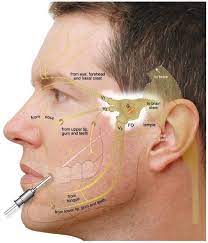
Trigeminal neuralgia (TN) is a debilitating neurological disorder that causes sudden and severe pain, usually on one side of the face. In this chronic condition, the trigeminal nerve, responsible for transmitting sensations from the face to the brain, becomes irritated or compressed, leading to sharp, shooting pain in the affected area.
There are three types of trigeminal neuralgia: classical, secondary, and idiopathic trigeminal neuralgia. Various factors contribute to this neurological condition, including nerve damage, ageing, inflammation, and compression of the trigeminal nerves.

Symptoms of Trigeminal Neuralgia
Some of the symptoms of trigeminal neuralgia include the following:
- Severe facial pain
- Regular aches and pains
- Numbness or a tingling sensation
- Burning sensation on one side of the face
- Short bursts of severe pain

Certain simple activities, such as touching your face, eating, and drinking, and applying any impact or pressure to your face, can trigger painful episodes.
Triggers for Trigeminal Neuralgia
The pain of trigeminal neuralgia can be triggered by various factors, including:
- Everyday activities involve movement of the facial muscles, such as chewing, talking, or smiling.
- Touch or vibrations include washing the face, applying makeup, or shaving.
- Exposure to cold air or wind on the face, especially around the mouth and nose.
- Eating and drinking
- Brushing your teeth, flossing, and using mouthwash
- Applying any impact or pressure to your face, especially to your cheek or jawline
Surgical Options for Trigeminal Neuralgia
While medications are often the first line of treatment for trigeminal neuralgia, some individuals may not achieve adequate pain relief or may experience undesirable side effects. In such cases, surgical intervention may be considered to alleviate pain and improve the overall well-being of the patient.
Some of the surgical procedures for trigeminal neuralgia treatment are discussed below.
- Microvascular Decompression (MVD)

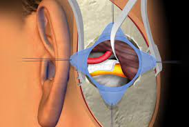
It is one of the major surgical options for treating trigeminal neuralgia, as it is a less invasive surgical procedure. During the procedure, the neurosurgeon places a tiny cushioning pad or Teflon sponge between the trigeminal nerve and the blood vessels, effectively insulating the nerve from contact. MVD has shown excellent long-term results in reducing or eliminating pain in many patients.
2. Radiofrequency Rhizotomy

It is a minimally invasive procedure that is used to remove sensation from a painful nerve that is sending pain signals to the brain. This procedure is carried out by inserting a needle through the cheek, which is then used by surgeons to send electric signals to identify the specific pain point on the trigeminal nerve. Radiofrequency rhizotomy is also known as radiofrequency ablation, in which a radiofrequency current is used to burn the fibres.
3. Glycerol Injection Rhizotomy

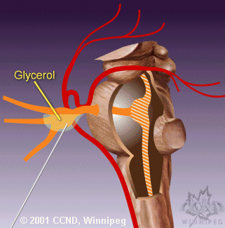
Glycerol injection is a less invasive alternative to surgical procedures that involves injecting glycerol, a chemical agent, into the trigeminal nerve root. The glycerol causes the nerve to compress and damage, leading to pain relief.
CONCLUSION
For those suffering from the relentless pain of trigeminal neuralgia, surgical options provide significant pain relief and improved quality of life. Individuals with trigeminal neuralgia must work closely with their neurosurgeon to explore the most suitable surgical option based on their unique medical history and condition.
With advancements in neurosurgical techniques, many patients can find lasting relief from the burden of trigeminal neuralgia and rediscover a life free from debilitating pain.
Consult a neurosurgeon to get an effective trigeminal neuralgia treatment plan according to your physical health and the severity of the symptoms.
A brain cyst is a fluid-filled sac or lesion that forms within the brain tissue or in its surrounding membranes. The cyst is mostly benign, which means it does not lead to any cancer but can affect the daily routine of an individual in ways such as headaches, vision problems, nausea and vomiting, seizures, and many others.
What Is an Intraventricular Brain Cyst?
The intraventricular cyst is a condition in which the development of cysts occurs within the brain ventricles.
Ventricles are the cavities in the brain that are responsible for the production and storage of cerebrospinal fluid. CSF is a watery fluid that surrounds your brain and spinal cord and also helps provide cushioning and protection from any trauma. It also removes waste and provides nutrients to your brain.
When a cyst develops in any of the ventricles, it can disrupt the cerebrospinal fluid’s normal flow and functioning, leading to various potential complications.

Colloid Cyst:
A colloid cyst is a type of brain cyst that typically develops in the third ventricle of the brain. The third ventricle is one of the four interconnected cavities within the brain responsible for cerebrospinal fluid (CSF) circulation. Colloid cysts are filled with a thick, gel-like substance called colloid material. While these cysts are generally considered benign, they can cause problems by obstructing the normal flow of cerebrospinal fluid.
Causes and Symptoms of an Intraventricular Brain Cyst
The causes of intraventricular brain cyst are still unknown and can vary from person to person. It is believed that some cysts are congenital, i.e., present at birth due to some fetal abnormalities, and others may be acquired later in life, like head injuries, inflammation within the brain, and infections.
Signs and symptoms of an intraventricular brain cyst include persistent headaches that worsen over time, blurred or double vision, changes in mood and behaviour, and difficult concentration and coordination issues.
Surgical Options for Intraventricular Cysts
Treatment options for intraventricular cysts are considered when the cyst in the ventricles may press the brain tissue and cause symptoms or when it becomes enlarged. Treatment is also recommended when the cyst blocks the normal flow of cerebrospinal fluid.
Neuroendoscopic options to treat your intraventricular cyst:
Neuroendoscopy is a minimally invasive surgical procedure that is used to treat intraventricular cysts that develop in the brain’s ventricles. Under neuroendoscopy, several treatments are classified based on the type, characteristics, and location of the cyst. It also depends on the patient’s overall health, and below are some of the surgical options that use neuroendoscopy to treat intraventricular cysts.

- Cyst fenestration
One of the best and most effective treatments for intraventricular cysts is endoscopic cyst fenestration. It is a minimally invasive procedure that includes creating an opening, also known as fenestration, in the cyst while using an endoscope.
During this surgical procedure, the surgeon inserts the endoscope into the ventricles to help doctors look out the cyst and carefully create an opening in the cyst. Creating the opening in the cyst, allows the cyst’s contents (cerebrospinal fluid) to drain into the surrounding ventricle or brain tissue.
2. Cyst resection
There are different types of intraventricular cysts, and so there are treatments. It is necessary to completely remove the solid and thick-walled intraventricular cyst.

For this, surgeons choose the neuroendoscopic cyst resection method. This surgical method involves using an endoscope to carefully visualise the cyst and dissect it from the surrounding tissue.
After removing the cyst, the patient will not experience any symptoms or complications associated with the cyst.
3. Endoscopic Third Ventriculostomy (ETV)
This procedure is performed when the intraventricular cyst interferes with the flow of the cerebrospinal fluid within the ventricles. During ETV, doctors, by using an endoscope, create an opening in the floor of the third ventricle that helps allow the fluid to bypass the cyst and resume its normal functioning.

Neuroendoscopy for intraventricular cysts offers several benefits such as less invasive, shorter recovery period, and hospital stays as compared to traditional surgery.
4. Endoscopic Excision of Colloid Cysts
One of the most advanced and minimally invasive methods for excising colloid cysts is through neuroendoscopy. Here’s how the procedure typically works:
- The patient is prepared for surgery, which may involve general anaesthesia or sedation.
- Endoscope Insertion: A neurosurgeon makes a small incision in the scalp and inserts a thin, flexible tube called an endoscope through a small burr hole in the skull. The endoscope has a light source and a camera that allows the surgeon to see inside the brain.
- Cyst Visualization: The surgeon navigates the endoscope to reach the location of the colloid cyst within the third ventricle. The camera provides a clear view of the cyst and surrounding structures.

- Cyst Resection: Using specialized instruments passed through the endoscope or by using laser technology, the surgeon carefully removes the colloid cyst and any obstructive material. The goal is to completely excise the cyst while minimizing damage to surrounding brain tissue.
- Closure: Once the cyst has been excised, the surgeon closes the incision, often with sutures or staples.
CONCLUSION
An intraventricular cyst is usually a benign cyst, but it can also lead to some discomfort and complications in individuals. To help alleviate the discomfort of people dealing with this problem, surgeons use specific approaches and techniques involving neuroendoscopy. It is important to consult a skilled neurosurgeon with extensive experience and specialising in neuroendoscopy.


